CellChek® D | D+
Designed to improve patient outcomes, CellChek® D+ redefined specular microscopy in Eyebanking.
It was quickly and widely adopted by many of the world’s finest Eye Banks.


CellChek® D+
Introducing the first multi-imaging system for donor corneal analysis. CellChek® D+ provides an amazing view into the cornea that simply has never been seen before defining the structure of the endothelium, stroma and epithelium. Comprehensive imaging to assist both eye banks and corneal surgeons to make better decisions on implantable material.
Now featured in Cornea June 2018 Supplement: Current and New Technologies in Corneal Donor Tissue Evaluation Comparative Image Atlas. Especially check out S7 “Wide-Field Ex Vivo Dual Imaging Microscopy.”
Clinical Applications
Clinical Benefits
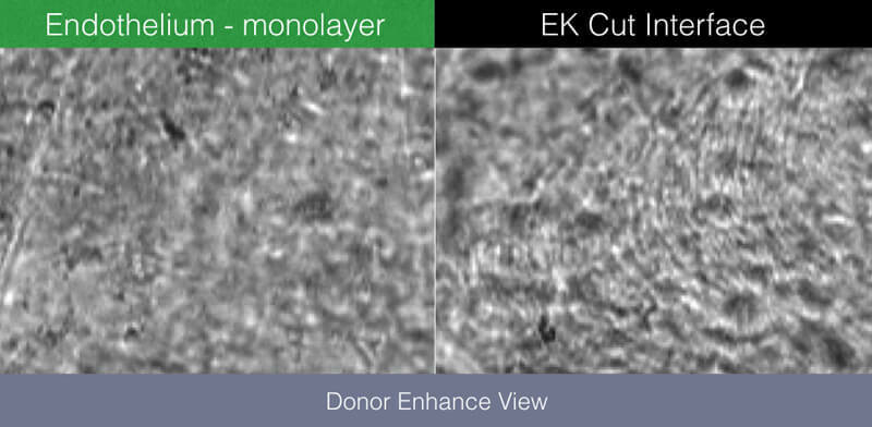
Provides remarkable views of endothelium, intra-stromal structure and the epithelium
For the first time, visualize blood cells, fungus, rough keratome cuts, dead cells, and epithelial anomalies
Regulatory
In Vitro USA

New “Donor Enhanced” Imaging System
CellChek® D+ features a patent applied for “Donor Enhance” imaging system that provides remarkable views of endothelium, intra-stromal structure, and the epithelium. The range of new information from which to make better clinical decisions is spectacular (pun intended): blood cells, fungus, rough keratome cuts, dead cells, epithelial anomalies. Never have we had this view of pre-implanted material. Corneal surgeons are finding this information to be illuminating in helping to understand graft success and failure.
Broad overview but with large multi-sample analytic areas
CellChek® D and D+ feature a new finder feature with digital measuring / documenting tools.


Patented Cellular Analysis Methods
All Konan specular microscopes feature the Center Method of analysis. Center Method is mentioned in FDA panel minutes as being the “gold standard” and is used by virtually every professional reading center for independent assessment of corneal endothelial analytics.¹ Unlike full-auto assessments, the Center Method provides high precision and repeatability for specular images in which relatively small continuous areas of visible cells are visible.
The Flex-Center method is the third tool for advanced stage diseased corneas in which only a very few number of cells are visible. With this semi-automated, perimeter-count method, again, high precision and repeatability is achieved. Only Konan provides the rich set of analytic tools for reliable assessment of the entire spectrum of corneal conditions.
It is not surprising that Konan is the global gold standard in corneal endothelial assessment.
Full Graft Imaging
CellChek® D and D+ provide a total picture of the cornea. Use digital measurement tools to identify, measure and document cornea dimensions, defects, and scars.

- Clinical and Scientific Publications
- Enhanced Donor Assessment
- Frequently Asked Questions
- Product Specifications
Clinical and Scientific Publications

As a corneal specialist for the last 22 years I would feel incomplete without my ability to perform specular in the clinical and eye bank setting. I have Konan units from the earliest to the most recent, all in service currently. They are reliable, accurate, and serve an essential role in my clinical practice and eye bank operations. The service delivered by Konan is some of the best I have experienced in the ophthalmic field. Specular microscopy provides a superb method of clinical screening for disease, an educational training tool for patients, their families and office and eye bank personnel, as well as a tissue evaluative method for eye banking. It is time and cost effective. I wouldn’t want to practice without a Konan specular microscope.
George O. Rosenwasser, MD

I have used Konan’s eye bank specular microscopes since November 2000. During that time we have processed over 45,000 corneas for transplant. Konan’s microscopes have been proven to be extremely reliable day in and day for a 24/7 operation.
The specular microscope is one of the most vital pieces of equipment for any eye bank. I have enjoyed working with Konan’s professional team as they are always readily available for any question or issue that may arise. I am looking forward to the new CellChek® D and CellChek® D+ which will be the new generation of donor cornea imaging & assessment.
Jason K. Woody
Enhanced Donor Assessment
Important New Endothelial Detail Discovered with CellChek® D+
Remarkable imaging of non-reflective “grey areas” for more definitive assessment.
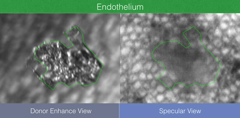
CellChek® D+ Provides Visualization of Non-Endothelial Features of the Posterior Cornea
View many features simply invisible to specular only donor imaging:
- Blood cells
- Scar tissue
- Debris
- Fungus
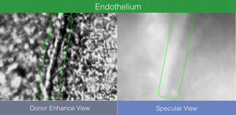
Image Intra-stromal Keratome and Femtosecond Laser Interface Quality with CellChek® D+
Interface features are invisible to specular-only donor systems.
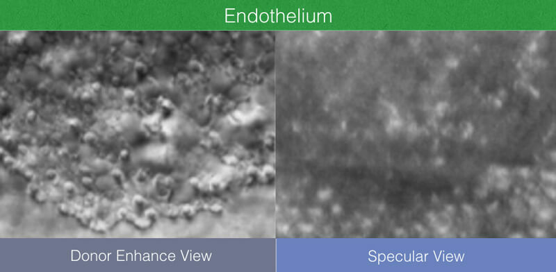
The Epithelium Now Revealed with CellChek® D+
View many features simply invisible to specular only donor imaging:
- Blood cells
- Scar tissue
- Debris
- Fungus
What is KonanCare support and clinical training?
How accurate are the analysis methods?
How is the system calibrated?
How many cells should be counted to be statistically accurate?
How do I set a focal distance between the objective lens and the endothelial layer using the different containers, such as vial or chamber?
What temperature range is required for corneal assessment (after removing from refrigeration)?
How is the imaging affected with cold corneas and how do we determine if the cornea has warmed up enough to analyze?
Product Specifications
CellChek® D+ |
EB-10 |
|
| Viewing Field | 1000 x 750 µm | 480 X 600 µm |
| Analysis Area (each multi-field area) | 400 X 300 µm X 4 areas (max 480,000 µm2) | 200 X 280 µm |
| Chamber Compatibility | Numedis Life 4C®, Krolman®, B+L®, Stephens® | Life 4C®, Alcon®, Bausch+Lomb®, Krolman® |
| Vial Dimensions | up to 35 mm diameter | up to 35 mm diameter |
| Thermometer Range | 0° to 45° C | 0° to 45° C |
| Stage Translation Ranges | X, Y, and Z = 16 mm, Tilt = 15° | X and Y = 16 mm, Z= 20 mm, Tilt: 5° |
| Illumination | Halogen | LED: primary wavelength 525 nm |
| Cameras | Dual CMOS: Finder and cornea | Dual CMOS: Finder and Cornea |
| Display | Digital data feed to computer | On-board LCD: temperature and pachymetry |
| Electrical | 100-240 VAC, 50/60 Hz, 50 VA | 100-240 VAC, 50/60 Hz, 50 VA |
| Size | 280 (W) X 215 (H) X 265 (D) mm | 200 (W) X 255 (H) X 220 (D) mm |
| Data Interface | USB 2.0 | USB 2.0 |
| Weight | 7.5 kg (without computer) | 6.1 kg (without computer) |
| Operating Conditions | Ambient temp: 10° to 40° C Relative humidity: 30% to 85% Atmospheric pressure: 70 to 106 kPa |
Ambient temp: 10° to 40° C Relative humidity: 30% to 85% Atmospheric pressure: 70 to 106 kPa |
| Regulatory | CE Mark, In Vitro USA | CE Mark, In Vitro USA |
What Konan Medical Customers Say

The service delivered by Konan is some of the best I have experienced in the ophthalmic field. Specular microscopy provides a superb method of clinical screening for disease, an educational training tool for patients, their families and office and eye bank personnel, as well as a tissue evaluative method for eye banking.
George O.D. Rosenwasser, MD
Central Pennsylvania Eye Institute – Medical Director, Gift of Life Donor Program Eye Bank

I have used Konan’s eye bank specular microscopes since November 2000. Konan’s microscopes have been proven to be extremely reliable day in and day for a 24/7 operation. I have enjoyed working with Konan’s professional team as they are always readily available for any question or issue that may arise. I am looking forward to the new CellChek® D and CellChek® D+ which will be the new generation of donor cornea imaging & assessment.
Jason K. Woody
President & CEO, Lions Eye Institute