CellChek®️️️ 20 & 20 PLUS
Blazing Fast, Fully-Automated Specular Microscopes
Process bilateral exams with one touch in about 40 seconds
Konan specular microscopes were recognized as the gold standard specular microscopes in the minutes from an FDA panel meeting*

The Gold Standard, Redefined.
Faster, Easier Endothelial Imaging
CellChek®️️️ 20 is Konan Medical’s newest, non-contact specular microscope that can capture and analyze bilateral exams with one touch in under 40 seconds.
The new easy-to-use “Simple Mode” enables one-touch, fully-automated endothelial image capture with analysis, reporting, and exporting of data.
WATCH THE REAL-TIME VIDEO >>>
Clinical Applications
Glaucoma, cataract, & refractive surgery.
Corneal disease management.
Contact/specialty contact lens fittings.
Routine eye care
Clinical Benefits
Visualize endothelial cells with 40x magnification compared to slit lamp bio-microscopy
Identify pre-existing low density and dystrophies that may affect positive surgical outcomes
Confirm recommended minimum cell density/morphology for scleral/specialty contact lenses
Regulatory
FDA 510(k) Cleared | CPT Code 92286
CE Marked
Health Canada Licensed

Blazing Fast
The new, easy-to-use “Simple Mode” enables one-touch, fully-automated endothelial image capture with analysis, reporting, and exporting of data.
The completely reimagined CellChek®️️️ 20 is noticeably improved and simplified; remove the device from the box, connect to power, turn it on, and start imaging.
Konan’s gold standard Center Method™ and Flex Center Method™ are now fully-automated. Automated Center Method displays cell center counted densities and morphometric indices of both eyes immediately after images are captured – all in under 40 seconds.
Key New Features
With CellChek®️️️ 20 you can capture, analyze and print/export a bilateral exam with just one touch. The new, easy-to-use “Simple Mode” makes running a bilateral exam faster and easier than ever before. CellChek®️️️ 20 reduces testing time significantly, allowing you to test patients quickly and efficiently.
One-touch imaging
Fast, one-touch imaging of both eyes including positioning, alignment, imaging capture, analysis and print-out/exporting data.
Optimized touch functions
Easy touch-friendly interface makes capturing, analysis, and reporting fast and efficient. Also features pinch-to-zoom image adjustment.
25% higher resolution
Improved resolution allows better visualization of single cells and cell walls. Superb cell border tracing and guttae exclusion.
Auto-capture/retry
This improved technology allows for successful imaging of difficult or challenging cases.
30% larger image area
The field of view has been increased to 0.25mm x 0.55mm, enabling better corneal visualization and more cells to be analyzed.
CellChek®️️️ 20 PLUS
CellChek®️️️ 20 PLUS adds near-limbal imaging to the standard model. It extends the limits of corneal endothelial image capture adding locations to 4.5mm from center. Six additional fixation points, 13 total.


Flexible Touch Screen
The flexible 10.6” wide touch screen can be turned and tilted 180°, providing better ergonomics, space savings, and flexibility with technician use from any of the four sides: front, back, left, and right.
Fully-Automated Analysis
CellChek®️️️ 20 offers fully-automated: Center Method; Flex-Center Method; and Auto-Trace analysis. These analysis methods provide one-touch, high precision results FAST.
These fully-automated analysis methods are offered alongside the traditional manual Center and Flex-Center analysis methods, offering a total of 6 analysis options.
Konan’s Center Method is mentioned in FDA panel minutes as being the “gold standard”, and is used by virtually every professional reading center.

New Auto Center Method
With the new Auto Center Method, the center of each individual, visible cell is automatically marked. Guttae and other dark regions are automatically excluded.
New Auto Flex-Center Method
With the new Auto Center Method, the center of each individual, visible cell is automatically marked. Guttae and other dark regions are automatically excluded.

Clinical Benefits
Cataract Surgery and
Premium IOLs
Low endothelial cell counts and pre-existing dystrophies can markedly reduce the potential for positive surgical outcomes from an otherwise uneventful cataract surgery. Surgeons are finding these data points critical when recommending premium IOLs: verify and document pre-operatively that the cornea is not suspect to more likely post-op complications difficult to explain with the investment in premium IOLs.
General Corneal Health Assessment
Konan specular microscopy is an invaluable tool to screen for corneal diseases as such as Fuchs’ Dystrophy, keratoconus, other corneal dystrophies, and trauma. You won’t believe what you’ve been missing.
Refractive Surgery
Contact Lenses
Testimonials
![]()

Central Pennsylvania Eye Institute – Medical Director, Gift of Life Donor Program Eye Bank
![]()

Solomon Eye Physicians & Surgeons, Bowie, Maryland – Greenbelt, MD, USA
![]()

Pacific ClearVision Institute
![]()

Devers Eye Institute, Portland, OR USA
![]()

Woolfson Eye Institute, Sandy Springs, GA, USA
![]()

Cleveland, OH USA
![]()

Director, Wang Vision 3D Cataract & LASIK Center Clinical Associate Professor of Ophthalmology, University of Tennessee International President, Shanghai Aier Eye Hospital
![]()

Eyecare of Greenville, Greenville, TX USA
![]()

Medical Director, Hazelton and Stroudsburg Eye Specialists, Hazelton, PA USA
![]()

Vision Source, Waco Vision and Health, Waco, TX USA
![]()

Contact Lens and Vision
![]()

Clinical Director, Kentucky Eye Institute
Chief Clinical Editor, Review of Optometry
Associate Professor, Kentucky College of Optometry
![]()

Gulf Coast Eye Center, Sarasota, FL USA
Publications
READ FULL ARTICLE
READ FULL ARTICLE
READ FULL ARTICLE
Fundamentals
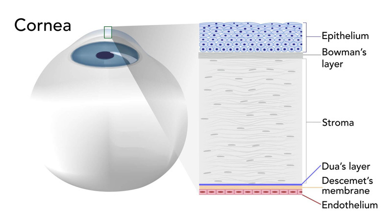
The Cornea
The corneal endothelium is a single layer of cells whose function is to maintain the balance of fluid (aqueous) within the cornea by means of a barrier effect, and to remove excess aqueous from the cornea by means of a pumping mechanism. A properly functioning endothelium maintains the correct clarity and shape of the corneal required for clear vision. When endothelial cells are lost or damaged, the remaining cells grow in size and change shape to fill in the gaps in order to maintain structural integrity. If too many cells are lost or damaged, the pumping mechanism may be negatively affected, resulting in corneal edema which may lead to partial or complete loss of vision.
Cell Density & Morphology Changes
Endothelial cells may be lost due to traumatic injury, damage during eye surgery (corneal incisions, phaco-energy), laser surgery, intraocular lenses, pharmaceutical agents contained in eye drops, or simply through the aging process. The number of cells along with the variance of the sizes and shapes of the endothelial cells serve as quantitative and qualitative indicators of the health of the cornea. With the prevalence of corneal and anterior segment surgeries, implantable eye devices, and contact lenses, the value of monitoring the corneal endothelium has never been higher. Konan’s specular microscope makes it possible to observe the endothelium at high magnification and provides a detailed assessment of the cornea. This important information helps define the best mode of surgery or therapy for a given patient.
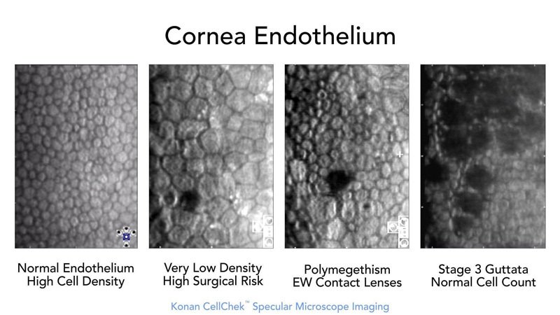
Endothelial Morphology Examples

Normal Endothelial Cell Density (ECD)
Low ECD
Polymegethism
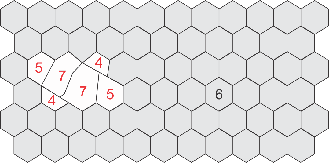
Pleomorphism
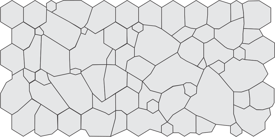
Normal ECD with Polymegethism and Pleomorphism
Konan Medical is trusted to provide the most widely used specular microscopy – endothelial analysis tools for routine clinical practice, clinical trials, professional reading centers, and the world’s #1 eye bank system for qualification of donor corneal tissue.
This is not a coincidence, it has been and is continuously earned… only from Konan.
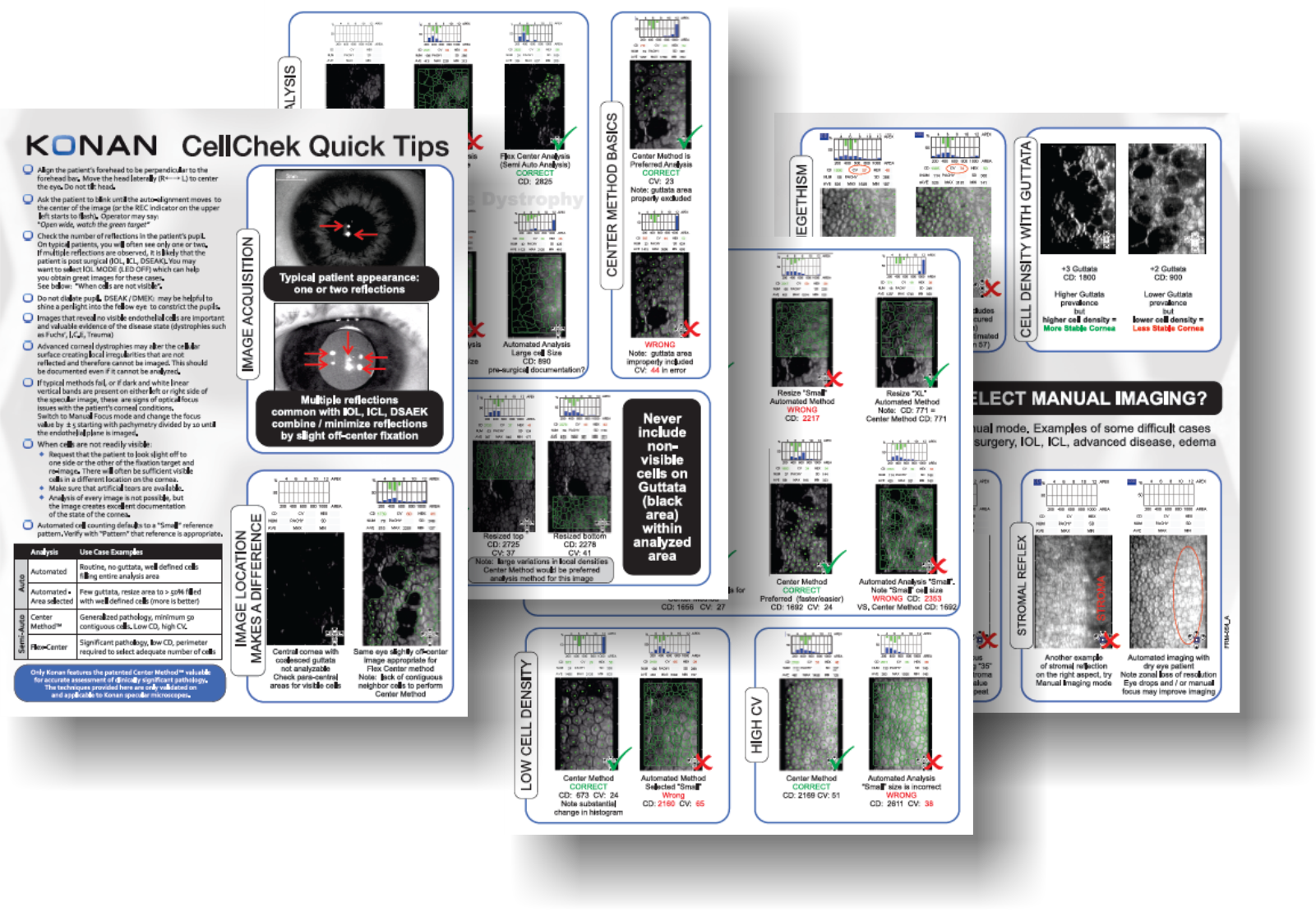
Konan CellChek®️️️ Quick Tips
Konan CellChek®️️️ system has long been the market leader for robust endothelial analysis. But it is the difficult cases where we really shine. Contact Konan to obtain a copy of CellChek®️️️ “Quick Tips” for in-depth clinical pearls for assessment of your most challenging cases including those that are good images of bad corneas.
ICL | PIOL | Scleral Lenses

Vance Thompson, MD

Theodore Perl, MD
It is important to select patients whose endothelial cell density and morphology are normal.
Avoiding Complications with Phakic IOLs” (Cataract and Refractive Surgery Today, October 2009)
ICL (implantable contact lens) or PIOL (phakic intraocular lens)
An endothelial cell count to verify that a patient’s cornea has an adequate endothelial cell density is an FDA labeling requirement for patient screening and selection prior to implantation of phakic intraocular lenses (Intraocular Contact Lenses, or “ICLs”). Without an adequate endothelial cell count, the current ICLs are contraindicated for implantation and clearly highlights the importance of this screening procedure. Endothelial cell density must be accurately checked preoperatively to ensure that a patient is a suitable candidate, and then should be monitored periodically postoperatively to verify the endothelium’s continued health. Although an endothelial cell count can be crudely estimated using a card developed in the 1980′s, this technique has significant limitations that are now well answered with Konan’s specular microscopy technologies:
- High precision and repeatability
- Excellent visualization of even stage +1 / +2 guttata that could significantly skew results
- Analysis of both cell counts and cell morphology changes (morphology is not assessed with a “card”)
- Changes in morphology can be a very sensitive indicator of corneal stress
- Unique ability to identify endothelial data location and assess trends over time
- Acquiring endothelial cell count or morphology changes over time from dissimilar locations can present erroneous assumptions on trends analysis
- Technician administered diagnostic test with full photographic documentation and statistics in only seconds.
- Does not require additional physician chair time and eliminates the inherent problems of “card” estimates.
- Important complementary uses for pre-operative assessments of corneal and other anterior segment procedures
ICL Products
Premium refractive products require premium attention to eligibility. Konan CellChek provides easy to use yet robust analytics to assure proper patient selection. There is a reason major manufacturers of ICL’s use Konan specular microscopes for collection of FDA safety data. Your patients deserve no less.

Visian ICLTM – Staar SurgicalTM

VerisyseTM – Abbott Medical OpticsTM

AcrySof® Cachet® – Alcon®
Visian ICL, Staar Surgical, Verisyse, Abbot Medical Optics, AcrySof Cachet, and ALCON are trademarks of their respective owners.
Scleral Lenses with High Risk Endothelial Cell Density
Patients with low endothelial cell densities may benefit from specular microscopy assessment to verify adequate density prior to fitting a scleral lens and to monitor continued supportive endothelial cell architecture if scleral lens treatments are implemented. Many experts recommend minimum cell densities of 800 to 1,000 cell / mm² to support scleral lenses.
Fundamentals of the importance of endothelial cell density to support implantation of phakic IOLs described above, may have parallels in decision making for scleral lens therapies.

Christine Sindt, OD, FAAO
Clinical Professor Ophtalmology and Vision Sciences, University of Iowa
It is common to experience corneal edema with endothelial cell counts under 800 cells/mm².

Eef van der Worp, BOptom, PhD, FAAO, FIACLE, FBCLE, FSLS
Pacific University, Oregon | University of Maastricht, Netherlands
Endothelial cell count of less than 800 cells/mm² is where the problems may arise (Sindt 2010a), and endothelial cell counts <1,000 cells/mm² should be handled with extra care and should not be fitted with scleral lenses to avoid edema.

Product Specifications
| Model no. / Product name | CellChek®️️️ 20-1 or CellChek®️️️ 20-1 PLUS / Konan Specular Microscope XX |
| Type | Class I, Type B |
| Photographic field | 0.25 x 0.55mm |
| Photographic capability | Automatic |
| Photographic location | Center, Peripheral locations (0, 45, 135, 180, 225, 315 degrees) |
| Central corneal thickness | Measurable |
| Illumination | LED |
| Analyzing method | Auto Center Method / Auto F Center Method / Auto Trace Method / Center Method / F Center Method / Trace Method |
| Analysis data | Cell density (mm2), Coefficient of Variation, Standard Deviation, Percentage of hexagonal cells, Numbers of calculated cells, Average cell area (µm2), Maximum cell area (µm2), Minimum cell area (µm2), Distribution of number of sides histogram (%), Distribution of area histogram (%) |
| Integrated monitor | 10.1” Capacitive touch panel with a 180 degrees flexible horizontal & vertical movement capability. |
| External interface | EMR connection / DICOM capability |
| External ports | USB3.0 x 4 Type A , RJ-45 |
| Power consumption | 100 VA |
| Dimensions | 310 (W) X 459 (D) X 451 (H) mm (with the monitor at the rear-side) |
| Weight | 19.6 kg |
What Konan Medical Customers Say

As a corneal specialist for the last 22 years I would feel incomplete without my ability to perform specular in the clinical and eye bank setting. I have Konan units from the earliest to the most recent, all in service currently. They are reliable, accurate, and serve an essential role in my clinical practice and eye bank operations. The service delivered by Konan is some of the best I have experienced in the ophthalmic field. Specular microscopy provides a superb method of clinical screening for disease, an educational training tool for patients, their families and office and eye bank personnel, as well as a tissue evaluative method for eye banking. It is time and cost effective. I wouldn’t want to practice without a Konan specular microscope.
George O. Rosenwasser, MD
Central Pennsylvania Eye Institute – Medical Director, Gift of Life Donor Program Eye Bank

Konan specular microscopes are recognized in the industry as the de facto standard for ECD determination. This is supported in the minutes from the FDA panel meeting to discuss the ARTISAN® lens: “Data from 12 sites were chosen because they used the Konan specular microscopes. This instrument is now the accepted standard for the most accurate determination of endothelial cell density.
R. Doyle Stulting, MD, PhD.

The Konan system takes specular microscopy to the next level, and allows for a swift and accurate appraisal of the most challenging corneas. All of my technicians are well versed in the use of the Konan specular microscope.
Jonathan D. Solomon, MD
Solomon Eye Physicians & Surgeons, Bowie, Maryland – Greenbelt, MD, USA

Simply an indispensable technology for Ophthalmology and Optometry, from Konan Medical, the “Apple” of ophthalmic device companies.
Paul Karpecki, OD, FAAO
SClinical Director, Kentucky Eye Institute Chief Clinical Editor, Review of Optometry Associate Professor, Kentucky College of Optometry