CellChek® D|D+
Frequently Asked Questions &
Specifications
Frequently Asked Questions
 KonanCare enables customers to consult, securely, by appointment, with Konan’s on-staff experts for interpretation of specular microscopic images, and to receive assistance on more advanced topics and methods associated with Konan’s products (CellChek XL, EvokeDx, CellChek D+ and EB-10, Chart2020, and the RAPDx pupillograph). With current product models, a secure, permission-based login session can be initiated by the customer allowing both the physician and the KonanCare team member to simultaneously view and discuss actual images using secure internet web services. KonanCare assistance is provided without charge for customers during the initial warranty period or during any period of Service Agreement coverage. A unique service, only from Konan Medical USA.
Konan's Center & Flex Center methods are patented and are the only FDA 510(k) cleared methods for eye bank systems. Accuracy established by independent reading centers for FDA clinical studies is +/-2.5%
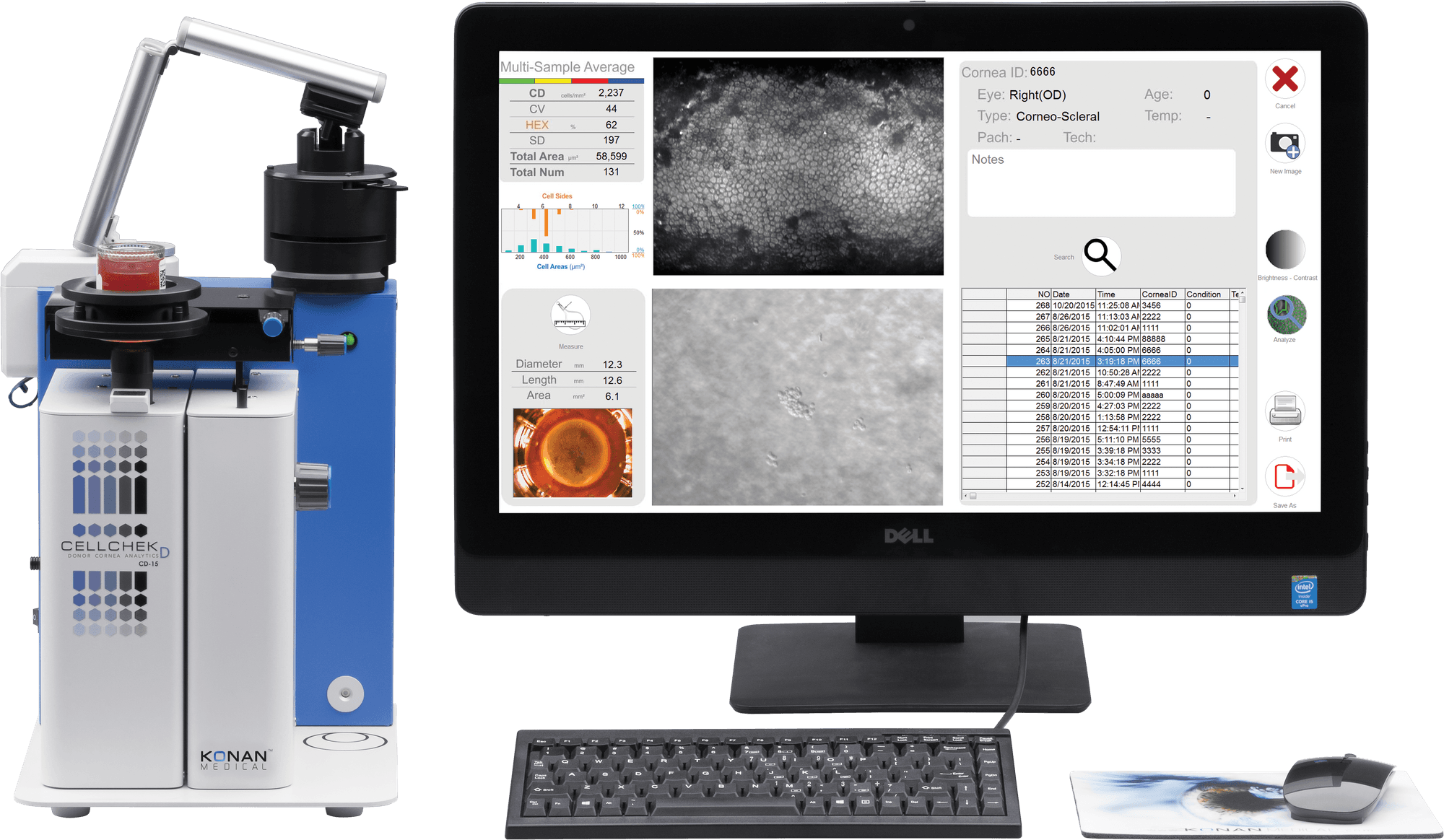
Each CellChek D+ comes with a 20um grid calibration disk. Enter 100um length using this disk and click a button. The computer will indicate a known magnification for 100um length. It also enables the operator to verify if calibration was properly completed.
Since it is difficult to determine the degree of cell size and shape variation in donor corneas, most eye banks are marking at least 75-100 cells per cornea. Konan’s multi-sampling capability enables the operator to choose up to 4 different locations in the central cornea for adequate sampling. Analysis from each frame can be automatically averaged.
Since the different containers place the cornea higher or lower than the focal point, by using the proper ring supplied for various containers, the endothelial layer is more or less focused when the top of the black stage and the top of the blue wall are at an even height. There is no need to use any ring for Krolman’s chamber.
CellChek D+ has a built-in thermometer which measures medium temperature in real-time. For optimal results, the preservation medium should be above 24 – 26 degrees C. (Remember that cornea is the happiest in the body-slightly less than 37 degrees C)
At 4º C, biochemical and physiological metabolic activities of endothelial cells slow down significantly. As temperature increases to ~ room temperature (>24º C), the endothelial cell's ability to pump, directly affects the image quality. If cornea has low functional reserve to start with, it may take longer for activation to occur.
Specifications
|
|
CellChek D+
|
EB-10 |
|
Viewing Field
|
1000 x 750 µm
|
480 X 600 µm
|
|
Analysis Area (each multi-field area)
|
400 X 300 µm X 4 areas (max 480,000 µm2)
|
200 X 280 µm
|
|
Chamber Compatibility
|
Numedis Life 4C®, Krolman®, B+L®, Stephens®
|
Life 4C®, Alcon®, Bausch+Lomb®, Krolman®
|
|
Vial Dimensions
|
up to 35 mm diameter
|
up to 35 mm diameter
|
|
Thermometer Range
|
0° to 45° C |
0° to 45° C
|
|
Stage Translation Ranges
|
X, Y, and Z = 16 mm, Tilt = 15°
|
X and Y = 16 mm, Z= 20 mm, Tilt: 5°
|
|
Illumination
|
Halogen
|
LED: primary wavelength 525 nm
|
|
Cameras
|
Dual CMOS: Finder and cornea
|
Dual CMOS: Finder and Cornea
|
|
Display
|
Digital data feed to computer
|
On-board LCD: temperature and pachymetry
|
|
Electrical
|
100-240 VAC, 50/60 Hz, 50 VA
|
100-240 VAC, 50/60 Hz, 50 VA
|
|
Size
|
280 (W) X 215 (H) X 265 (D) mm
|
200 (W) X 255 (H) X 220 (D) mm
|
|
Data Interface
|
USB 2.0
|
USB 2.0
|
|
Weight
|
7.5 kg (without computer)
|
6.1 kg (without computer)
|
|
Operating Conditions
|
Ambient temp: 10° to 40° C
Relative humidity: 30% to 85% Atmospheric pressure: 70 to 106 kPa |
Ambient temp: 10° to 40° C
Relative humidity: 30% to 85% Atmospheric pressure: 70 to 106 kPa |
|
Regulatory
|
CE Mark, In Vitro USA
|
CE Mark, In Vitro USA
|