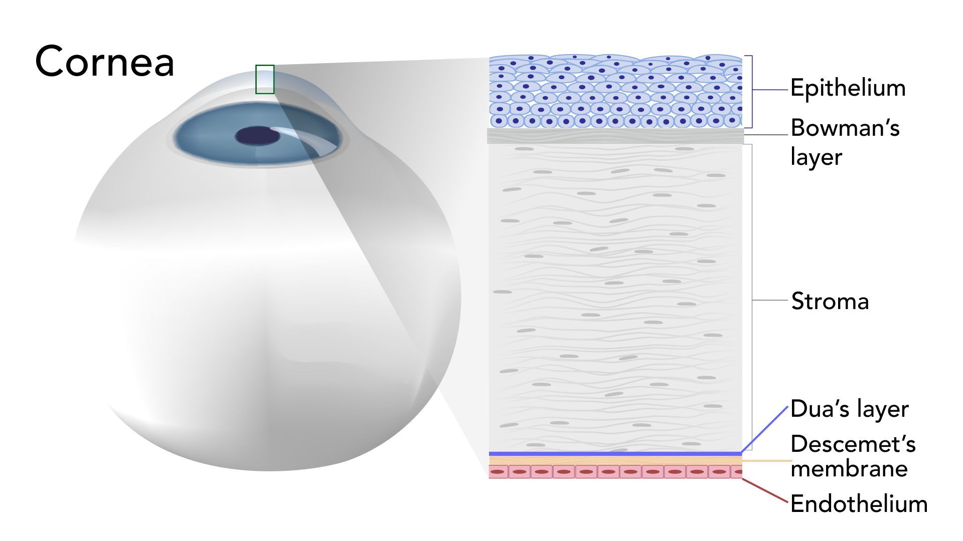
The Cornea
The corneal endothelium is a single layer of cells whose function is to maintain the balance of fluid (aqueous) within the cornea by means of a barrier effect, and to remove excess aqueous from the cornea by means of a pumping mechanism. A properly functioning endothelium maintains the correct clarity and shape of the corneal required for clear vision. When endothelial cells are lost or damaged, the remaining cells grow in size and change shape to fill in the gaps in order to maintain structural integrity. If too many cells are lost or damaged, the pumping mechanism may be negatively affected, resulting in corneal edema which may lead to partial or complete loss of vision.
Cell Density & Morphology Changes
Endothelial cells may be lost due to traumatic injury, damage during eye surgery (corneal incisions, phaco-energy), laser surgery, intraocular lenses, pharmaceutical agents contained in eye drops, or simply through the aging process. The number of cells along with the variance of the sizes and shapes of the endothelial cells serve as quantitative and qualitative indicators of the health of the cornea. With the prevalence of corneal and anterior segment surgeries, implantable eye devices, and contact lenses, the value of monitoring the corneal endothelium has never been higher. Konan’s specular microscope makes it possible to observe the endothelium at high magnification and provides a detailed assessment of the cornea. This important information helps define the best mode of surgery or therapy for a given patient.
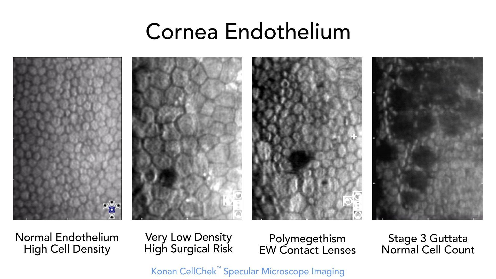
Endothelial Morphology Examples
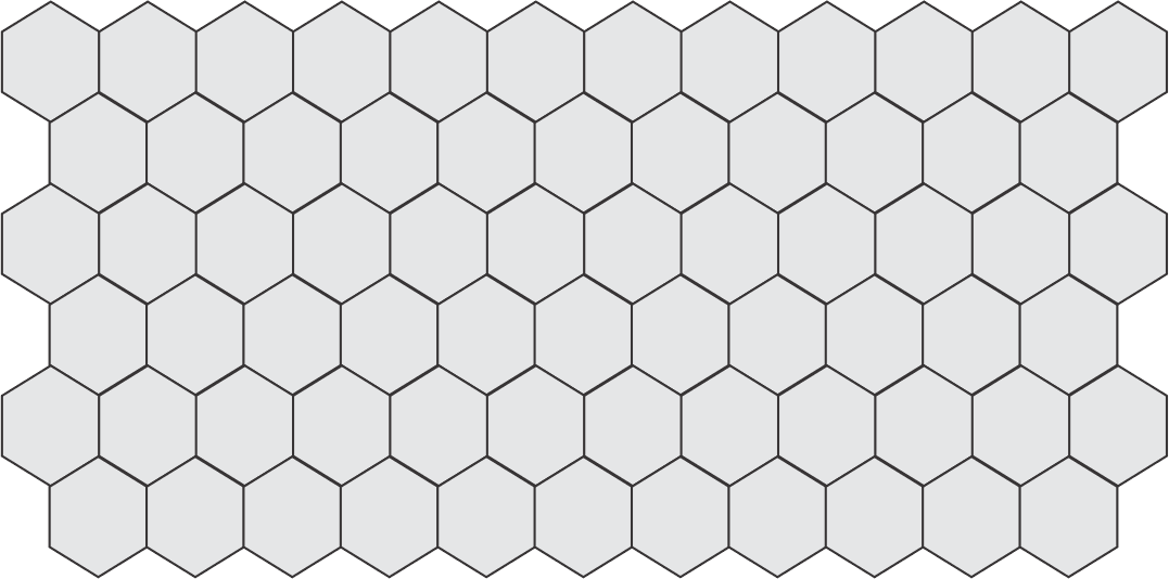
Normal Endothelial Cell Density (ECD)
Normal ECD declines with age.
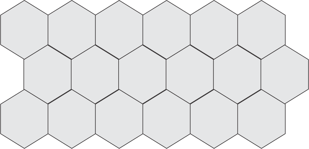
Low ECD
Low ECD is a risk factor for cataract and corneal surgeries that can be missed without the use of specular microscopy.
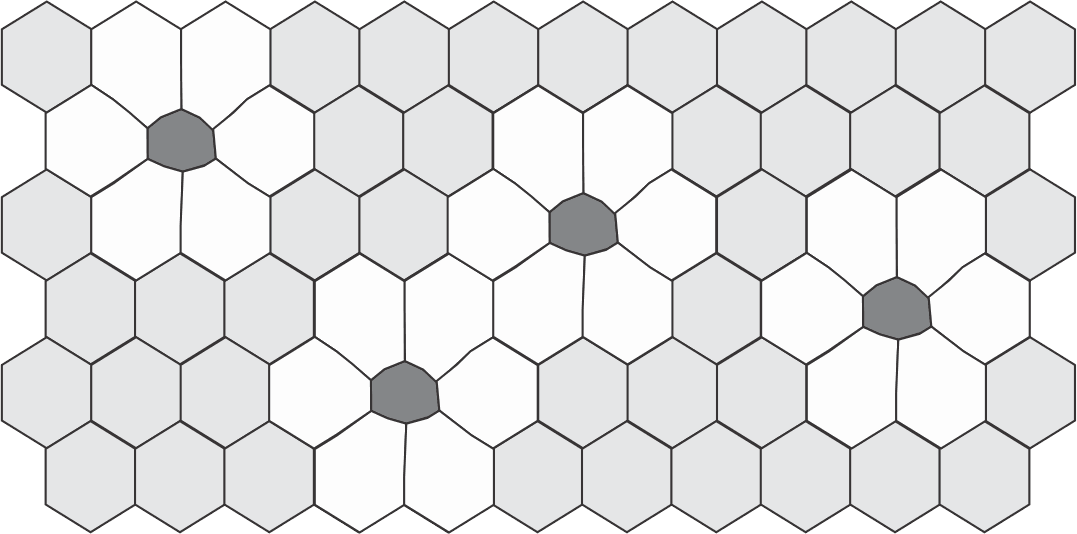
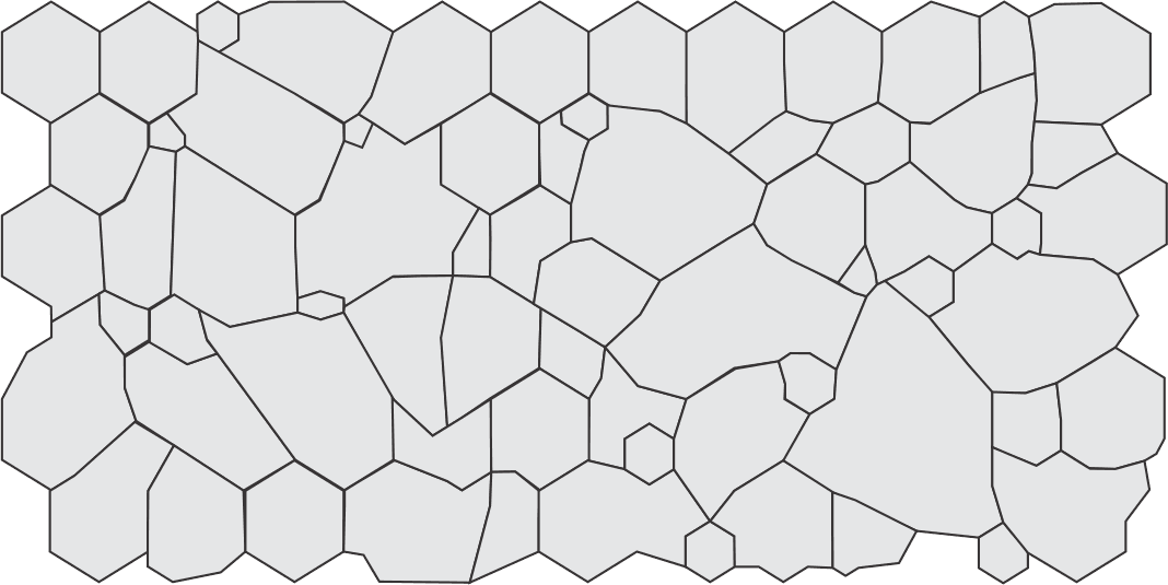
Polymegethism
Change from uniform cell sizes to variable cell sizes is an indication of distress to the endothelium.
Pleomorphism
Change from optimum hexagonal cell geometry to variable / compromised cell shapes.
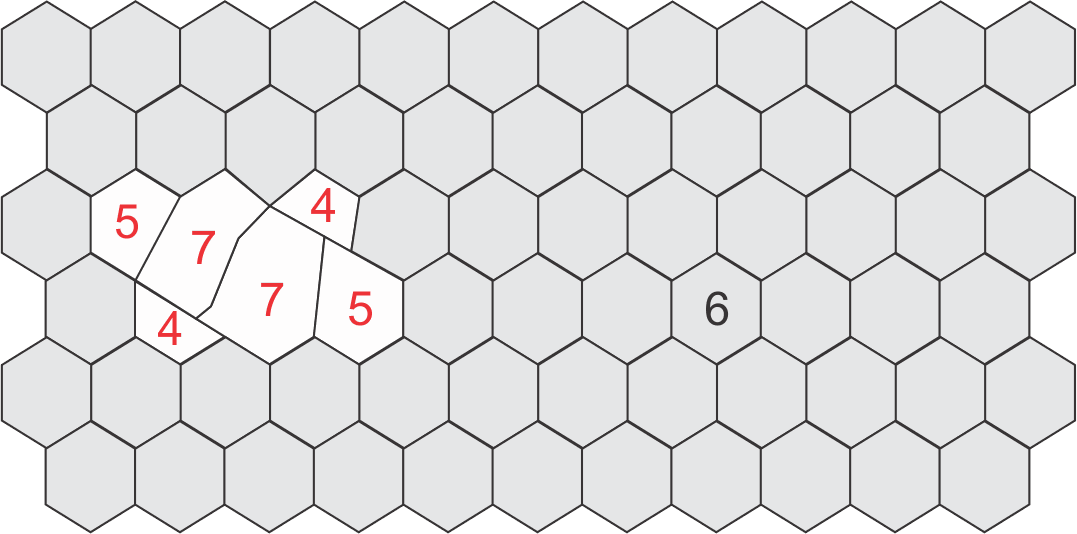
Normal ECD with Polymegethism and Pleomorphism
Here the ECD is typical, but there are also large changes in morphology with high variance in cell area / sizes (pleomorphism) and a large number of non-hexagonal cell shapes (4, 5, 7 sided cells, i.e. pleomorphism).
Konan Medical is trusted to provide the most widely used specular microscopy – endothelial analysis tools for routine clinical practice, clinical trials, professional reading centers, and the world’s #1 eye bank system for qualification of donor corneal tissue.
This is not a coincidence, it has been and is continuously earned… only from Konan.
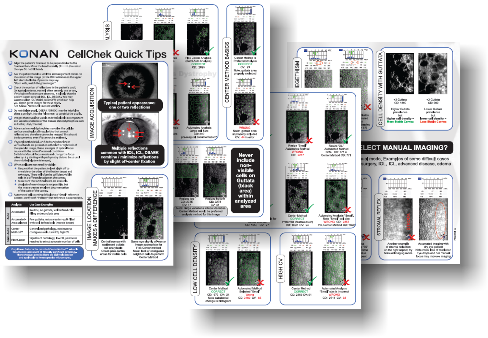
Konan CellChek Quick Tips
Konan CellChek system has long been the market leader for robust endothelial analysis. But it is the difficult cases where we really shine. Contact Konan to obtain a copy of CellChek “Quick Tips” for in-depth clinical pearls for assessment of your most challenging cases including those that are good images of bad corneas.