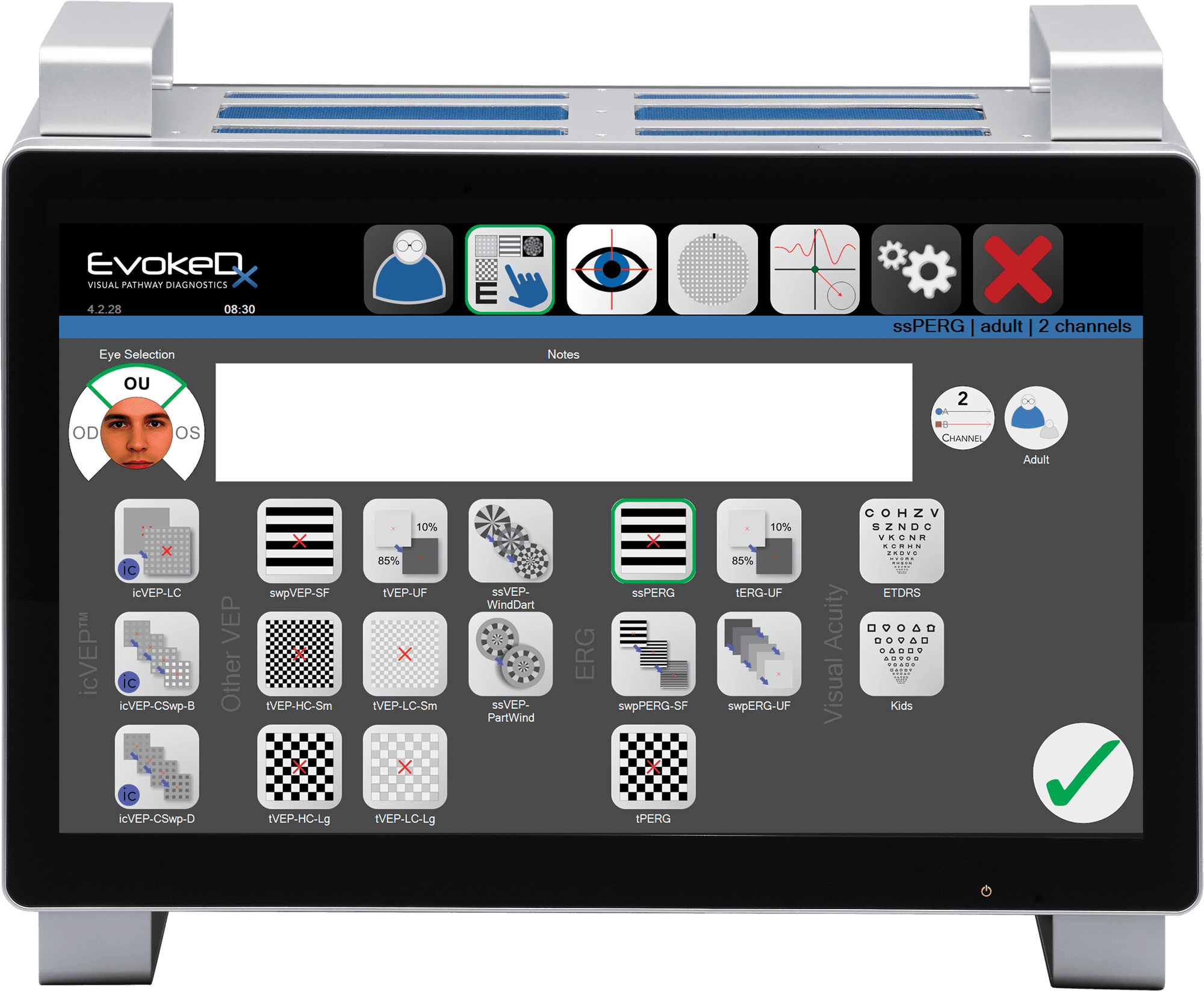

Konan’s EvokeDx is the most advanced electrodiagnostic device for use by eye care professionals I’ve ever seen. The myriad of testing options allows for the quantification of aspects of visual performance beyond anything else on the market. With more than 40 years using visual electrophysiology using many of the systems available, I really appreciate the new Fourier analytics methods of analyzing the data. It took a short time to wrap my head around, but the reward has been enormous.
Additionally, the gaze tracking system allows continuous monitoring of the patient’s attention and the OLED screen with all its concordant benefits of clean stimuli and precise control of contrast is suburb. EvokeDx is a winner!
Paul Harris, OD
Professor, Southern College of Optometry

As a next generation VEP and ERG system, EvokeDx provides greater ease of use and acquisition, a sleek portable high-tech system, a precise assessment of the full visual pathway with specific testing capabilities for important M cell pathways that aid in the accuracy of a diagnosis.
The system is touch screen is easy for the staff and the valuable information or readouts provided makes it easy for the doctor to assess pathology. Konan has become the “Mac” of ophthalmic technology and has once again done it with VEP and ERG testing..
Paul Karpecki, OD, FAAO
Clinical Director, Kentucky Eye Institute
Chief Clinical Editor, Review of Optometry
Associate Professor, Kentucky College of Optometry
The EvokeDx system includes a range of tests based on scientific findings accrued over decades that have elucidated the neural origins of the electrophysiological responses elicited noninvasively from the visual system. The systems analysis approach used involves quantitative analysis of the entire responses (VEPs/ERGs) in the frequency domain and their relations to time-domain waveforms.
Statistical methods applied to these measures enable determination of whether responses are present in the recordings and significance tests to compare responses from fellow eyes as well as changes over time associated with disease or treatments.
These techniques have been employed successfully to characterize contributions to the responses from and the integrity of different pathways and mechanisms.
Vance Zemon, PhD
Co-inventor EvokeDx | NeucodiaProfessor of Psychology, Ferkauf Graduate School Yeshiva University Albert Einstein College of Medicine
Clinical Resources
Accuracy of isolated-check visual evoked potential technique for diagnosing primary open angle glaucoma
Xu, L.J., Zhang, L., Li, S.L. et al. Doc Ophthalmol (2017). doi:10.1007/s10633-017-9598-6
Diagnostic performance of isolated-check visual evoked potential versus retinal ganglion cell-inner plexiform layer analysis in early primary open-angle glaucoma
Chen X, Zhao Y. Diagnostic performance of isolated-check visual evoked potential versus retinal ganglion cell-inner plexiform layer analysis in early primary open-angle glaucoma. BMC ophthalmology. 2017 Dec;17(1):77.
Novel electrophysiological instrument for rapid and objective assessment of magnocellular deficits associated with glaucoma
Zemon, Vance, et al. “Novel electrophysiological instrument for rapid and objective assessment of magnocellular deficits associated with glaucoma.” Documenta ophthalmologica 117.3 (2008): 233-243.
READ FULL ARTICLEInfluence of pupil size and other test variables on visual function assessment using visual evoked potentials in normal subjects
Salim, S., Childers, K., Lupinacci, A. P., Hu, G. Z., Zemon, V., & Netland, P. A. (2010). Influence of pupil size and other test variables on visual function assessment using visual evoked potentials in normal subjects. Documenta ophthalmologica, 121(1), 1-7.
READ FULL ARTICLEVisual evoked potential assessment of the effects of glaucoma on visual subsystems
Greenstein, Vivienne C., et al. “Visual evoked potential assessment of the effects of glaucoma on visual subsystems.” Vision research 38.12 (1998): 1901-1911.